 |
| HISTORY OF CT SCAN |
HISTORY OF CT SCAN
The Computed Tomography (CT) scan, also known as
a CAT (Computerized Axial Tomography) scan, is a medical imaging technique that
provides detailed cross-sectional images of the body. The history of the CT scan
dates back to the early 20th century when scientists and researchers were exploring
ways to visualize internal body structures without invasive procedures. The development
of the CT scan involved several key milestones:
- Theoretical Concepts (early 1900s): The theoretical
foundations of CT scanning were laid down by the Austrian mathematician Johann
Radon in the early 20th century. He developed the mathematical principles for
reconstructing an image from a series of X-ray projections, which laid the
groundwork for future developments in tomographic imaging.
- First Experimental CT Scanner (1963): The first
practical CT scanner was invented by British engineer Sir Godfrey Hounsfield
in 1963. Hounsfield was working at the EMI Laboratories in England when he
developed the first prototype. His revolutionary idea involved using X-ray
beams and detectors to collect multiple data points from different angles around
the patient's body. He then applied mathematical algorithms to reconstruct
the data into cross-sectional images.
- Introduction of Clinical CT Scanning (1971):
In 1971, the first commercial CT scanner, the EMI-Scanner (also known as the
"Mark I"), was installed at Atkinson Morley Hospital in London. The
scanner was used to image the brain and quickly proved to be a valuable diagnostic
tool in the medical field.
- Rapid Advancements and Widespread Adoption
(1970s-1980s): CT technology rapidly advanced during the 1970s and 1980s. Improvements
in computer processing power and imaging techniques led to faster scanning
times and higher image resolution. The technology spread worldwide, becoming
an indispensable tool for medical diagnosis and revolutionizing medical imaging.
- Development of Spiral CT (1989): In 1989, the
first spiral CT scanner was introduced by Siemens, which allowed continuous
volume scanning with faster acquisition times and improved image quality. Spiral
CT significantly enhanced the ability to image organs and blood vessels, making
it especially valuable in diagnosing conditions like cardiovascular disease.
- Multislice CT Scanners (1998): The introduction
of multislice CT scanners in 1998 further improved imaging capabilities. These
scanners could acquire multiple slices of the body simultaneously, reducing
scan times and enhancing image clarity.
- Evolution to High-Resolution and Cone Beam
CT (2000s): CT technology continued to evolve, with the development of high-resolution
CT scanners and cone beam CT scanners. These advancements expanded applications
in various medical specialties, such as oncology, orthopedics, and dentistry.
Today, CT scanning is a standard imaging tool used
in medical settings worldwide. Its ability to provide detailed cross-sectional images
has greatly improved the diagnosis and treatment of various medical conditions,
making it a vital component of modern healthcare.
CT scan
A computed tomography scan (usually abbreviated to CT scan; formerly called computed axial tomography scan or CAT scan) is a medical imaging technique used to obtain detailed internal images of the body. The personnel that perform CT scans are called radiographers or radiology technologists.
CT scanners use a rotating X-ray tube
and a row of detectors placed in a gantry to measure X-ray attenuations by different
tissues inside the body. The multiple X-ray measurements taken from different angles
are then processed on a computer using tomographic reconstruction algorithms to
produce tomographic (cross-sectional) images (virtual "slices") of a body.
CT scan can be used in patients with metallic implants or pacemakers, for whom magnetic
resonance imaging (MRI) is contraindicated.
Since its development in the 1970s,
CT scanning has proven to be a versatile imaging technique. While CT is most prominently
used in medical diagnosis, it can also be used to form images of non-living objects.
The 1979 Nobel Prize in Physiology or Medicine was awarded jointly to South African-American
physicist Allan MacLeod Cormack and British electrical engineer Godfrey Hounsfield
"for the development of computer-assisted tomography".
Types
On the basis of image acquisition and
procedures, various types of scanners are available in the market.
Sequential CT
Sequential CT also known as step-and-shoot
CT is a type of scanning method in which the CT table moves stepwise. The table
increments to a particular location and then stops which is followed by the X-ray
tube rotation and acquisition of a slice. Then the table increments again and another
slice is taken. The table has to make stop taking slices. This results in an increased
time of scanning.
Spiral CT
Drawing of CT fan beam and patient in a CT imaging system
CT scan of the thorax. The axial slice (right) is the image
that corresponds to number 33 (left).
Spinning tube, commonly called spiral
CT, or helical CT, is an imaging technique in which an entire X-ray tube is spun
around the central axis of the area being scanned. These are the dominant type of
scanners on the market because they have been manufactured longer and offer a lower
cost of production and purchase. The main limitation of this type of CT is the bulk
and inertia of the equipment (X-ray tube assembly and detector array on the opposite
side of the circle) which limits the speed at which the equipment can spin. Some
designs use two X-ray sources and detector arrays offset by an angle, as a technique
to improve temporal resolution.
Electron beam tomography
Electron beam tomography (EBT) is a
specific form of CT in which a large enough X-ray tube is constructed so that only
the path of the electrons, traveling between the cathode and anode of the X-ray
tube, are spun using deflection coils. This type had a major advantage since sweep
speeds can be much faster, allowing for less blurry imaging of moving structures,
such as the heart and arteries. Fewer scanners of this design have been produced
when compared with spinning tube types, mainly due to the higher cost associated
with building a much larger X-ray tube and detector array and limited anatomical
coverage.
Dual Energy CT
Dual Energy CT also known as Spectral
CT is an advancement of Computed Tomography in which two energies are used to create
two sets of data. A Dual Energy CT may employ a Dual source, Single source with dual
detector layer, and Single-source with energy switching methods to get two different
sets of data.
1. Dual-source CT is an advanced scanner with a two X-ray tube detector system,
unlike conventional single tube systems. These two detector systems are mounted
on a single gantry at 90° in the same plane. Dual Source CT scanners allow fast scanning
with a higher temporal resolution by acquiring a full CT slice in only half a rotation.
Fast imaging reduces motion blurring at high heart rates and potentially allows
for shorter breath-hold time. This is particularly useful for ill patients having
difficulty holding their breath or being unable to take heart-rate-lowering medication.
2. Single Source with Energy switching is another mode of Dual-energy CT
in which a single tube is operated at two different energies by switching the energies
frequently.
CT perfusion imaging
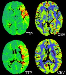
CT perfusion imaging is a specific form of CT to assess flow through blood vessels whilst injecting a contrast agent. Blood flow, blood transit time, and organ blood volume can all be calculated with reasonable sensitivity and specificity. This type of CT may be used on the heart, although sensitivity and specificity for detecting abnormalities are still lower than for other forms of CT. This may also be used on the brain, where CT perfusion imaging can often detect poor brain perfusion well before it is detected using a conventional spiral CT scan. This is better for stroke diagnosis than other CT types.
PET CT
PET-CT scan of the chest
Positron emission tomography-computed
tomography is a hybrid CT modality that combines, in a single gantry, a positron
emission tomography (PET) scanner and an x-ray computed tomography (CT) scanner,
to acquire sequential images from both devices in the same session, which are combined
into a single superposed (co-registered) image. Thus, functional imaging obtained
by PET, which depicts the spatial distribution of metabolic or biochemical activity
in the body can be more precisely aligned or correlated with anatomic imaging obtained
by CT scanning.
PET-CT gives both anatomical and functional
details of an organ under examination and is helpful in detecting different types
of cancers.
Medical use
Since its introduction in the 1970s,
CT has become an important tool in medical imaging to supplement conventional X-ray
imaging and medical ultrasonography. It has more recently been used for preventive
medicine or screening for disease, for example, CT colonography for people with
a high risk of colon cancer, or full-motion heart scans for people with a high risk
of heart disease. Several institutions offer full-body scans for the general population
although this practice goes against the advice and official position of many professional
organizations in the field primarily due to the radiation dose applied.
The use of CT scans has increased dramatically
over the last two decades in many countries. An estimated 72 million scans were
performed in the United States in 2007 and more than 80 million in 2015.
Head
Computed tomography of the human brain, from the base of the skull to top. Taken with intravenous contrast medium.
Commons: Scrollable computed tomography images of a normal
brain
CT scanning of the head is typically
used to detect infarction (stroke), tumors, calcifications, hemorrhage, and bone
trauma. Of the above, hypodense (dark) structures can indicate edema and infarction,
hyperdense (bright) structures indicate calcifications and hemorrhage, and bone
trauma can be seen as disjunction in bone windows. Tumors can be detected by the
swelling and anatomical distortion they cause, or by surrounding edema. CT scanning
of the head is also used in CT-guided stereotactic surgery and radiosurgery for the treatment of intracranial tumors, arteriovenous malformations, and other surgically
treatable conditions using a device known as the N-localizer.
Neck
Contrast CT is generally the initial
study of choice for neck masses in adults. CT of the thyroid plays an important
role in the evaluation of thyroid cancer. CT scan often incidentally finds thyroid
abnormalities, and so is often the preferred investigation modality for thyroid
abnormalities.
Lungs
A CT scan can be used for detecting
both acute and chronic changes in the lung parenchyma, the tissue of the lungs.
It is particularly relevant here because normal two-dimensional X-rays do not show
such defects. A variety of techniques are used, depending on the suspected abnormality.
For evaluation of chronic interstitial processes such as emphysema, and fibrosis,
thin sections with high spatial frequency reconstructions are used; often scans
are performed both on inspiration and expiration. This special technique is called
high-resolution CT produces a sampling of the lung and not continuous images.
HRCT images of a normal thorax in axial, coronal, and sagittal planes, respectively.
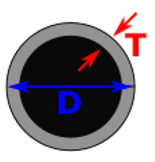
Bronchial wall thickness (T) and diameter of the bronchus (D)
Bronchial wall thickening can be seen
on lung CTs and generally (but not always) implies inflammation of the bronchi.
An incidentally found nodule in the
absence of symptoms (sometimes referred to as an incidentaloma) may raise concerns
that it might represent a tumor, either benign or malignant. Perhaps persuaded by
fear, patients and doctors sometimes agree to an intensive schedule of CT scans,
sometimes up to every three months and beyond the recommended guidelines, in an
attempt to do surveillance on the nodules. However, established guidelines advise
that patients without a prior history of cancer and whose solid nodules have not
grown over a two-year period are unlikely to have any malignant cancer. For this
reason, and because no research provides supporting evidence that intensive surveillance
gives better outcomes, and because of risks associated with having CT scans, patients
should not receive CT screening in excess of those recommended by established guidelines.
Angiography
Example of a CTPA, demonstrating a saddle embolus (dark horizontal line) occluding the pulmonary arteries (bright white triangle)
Computed tomography angiography (CTA)
is a type of contrast CT to visualize the arteries and veins throughout the body.
This ranges from arteries serving the brain to those bringing blood to the lungs,
kidneys, arms, and legs. An example of this type of exam is CT pulmonary angiogram
(CTPA) used to diagnose pulmonary embolism (PE). It employs computed tomography
and an iodine-based contrast agent to obtain an image of the pulmonary arteries.
Cardiac
A CT scan of the heart is performed
to gain knowledge about cardiac or coronary anatomy. Traditionally, cardiac CT scans
are used to detect, diagnose, or follow up coronary artery disease. More recently
CT has played a key role in the fast-evolving field of transcatheter structural
heart interventions, more specifically in the transcatheter repair and replacement
of heart valves.
The main forms of cardiac CT scanning
are:
- Coronary CT angiography (CCTA): the use of CT to assess the coronary arteries of the heart. The subject receives an intravenous injection of radiocontrast, and then the heart is scanned using a high-speed CT scanner, allowing radiologists to assess the extent of occlusion in the coronary arteries, usually to diagnose coronary artery disease.
- Coronary CT calcium scan: also used for the assessment of severity of coronary artery disease. Specifically, it looks for calcium deposits in the coronary arteries that can narrow arteries and increase the risk of a heart attack. A typical coronary CT calcium scan is done without the use of radiocontrast, but it can possibly be done from contrast-enhanced images as well.
To better visualize the anatomy, post-processing
of the images is common. The most common are multiplanar reconstructions (MPR) and volume
rendering. For more complex anatomies and procedures, such as heart valve interventions,
a true 3D reconstruction or a 3D print is created based on these CT images to gain
a deeper understanding.
Abdomen and pelvis
CT scan of a normal abdomen and pelvis, in sagittal plane, coronal and axial planes, respectively.
CT is an accurate technique for diagnosis
of abdominal diseases like Crohn's disease, GIT bleeding, and diagnosis and staging
of cancer, as well as follow-up after cancer treatment to assess response. It is
commonly used to investigate acute abdominal pain.
Non-enhanced computed tomography is
today the gold standard for diagnosing urinary stones. The size, volume, and density
of stones can be estimated to help clinicians guide further treatment; size is especially
important in predicting the spontaneous passage of a stone.
Axial skeleton and extremities
For the axial skeleton and extremities,
CT is often used to image complex fractures, especially ones around joints, because
of its ability to reconstruct the area of interest in multiple planes. Fractures,
ligamentous injuries, and dislocations can easily be recognized with a 0.2 mm resolution.
With modern dual-energy CT scanners, new areas of use have been established, such
as aiding in the diagnosis of gout.
Biomechanical use
CT is used in biomechanics to quickly
reveal the geometry, anatomy, density, and elastic moduli of biological tissues.


0 Comments